ORIGINAL ARTICLES
VOLUME: 20
ISSUE: 1
P: 28-35#28-35
March 2026
Acute Appendicitis and Enterobiasis: More Than A Coincidence?
Journal of Pediatric Infection • 2026
DOI: 10.5578/ced.20260118
Received Date: 02.12.2024
Accepted Date: 10.05.2026
Publish Date: 17.03.2026
ABSTRACT
Objective
Acute appendicitis (AA) and enterobiasis are common
problems of the childhood. The relationship between two incidents
have been controversial. The purpose of this study was to document
the rate and features of enterobiasis in paediatric appendicitis cases
of our hospital.
Material and Methods
This study was conducted in a tertiary
healthcare center. Data of 15 years were presented. The study and
control groups consisted of AA cases associated with and without
enterobiasis were matched according to age. The information on sociodemographic, clinical, laboratory, histopathological, and radiological
results were collected from on call surveys and hospital record system.
The statistical analyses were performed by statistical package program
software.
Results
The rate of enterobiasis was 1.6% (n= 53). Lower ultrasonographic appendiceal diameter, acute phase response and higher eosinophil levels were the features of the study group. Lymphoid hyperplasia was the most frequent histopathological diagnose.
Conclusion
AA is one of the most common surgical problems in paediatric emergency, but it is rarely associated with enterobiasis. History
of pruritus ani, lower signs of systemic inflammation, ultrasonographic diameter of the appendix and eosinophilia can warn the surgeon
about the parasite. Antihelminthic drugs are necessary to complete
the therapeutic process.
KEYWORDS
Enterobiasis, acute appendicitis, reactive lymphoid hyperplasia, children
INTRODUCTION
Acute appendicitis (AA), inflammation of the appendix due
to luminal obstruction, is one of the most common problems of
emergency services, requiring surgical intervention worldwide
(1,2).The incidence rate peaks in adolescence and females (3).It
is presented with sudden, unremitting abdominal pain, nausea,
vomiting, poor general condition, dehydration, tenderness in
the right iliac fossa, defence, and rebound (1).Although there
are many laboratory and radiological diagnostic facilities for
evaluation, all should be supported by repeating physical
examination and confirmed by histopathology (4). Luminal
obstruction is the pathophysiological reason for AA. Thus,
lymphatic drainage fails, and edema and mucosal ulceration
disrupt venous circulation. Ischemic necrosis occurs at the wall
of the appendix (5,6). Fecal stasis, fecaloids, tumors, bacterial,
parasitic, or protozoa infections may trigger this process.
Enterobiasis is one of the most popular unusual findings of
appendectomy specimens (7).
Enterobius vermicularis (E. vermicularis; pinworm, tapeworm,
threadworm, seatworm, oxyuriasis) is an obligatory nematode
and responsible for the most frequent gastrointestinal
parasitic infection in humans (8,9). It has a worldwide
distribution affecting all population groups and geographic
areas with higher incidence in low socioeconomic classes and
warm climate belts. It is more common in children and people
who live in close quarters with poor hygienic conditions (8-
10). Examining the Enterobius ova under the microscope via
cellophane tape or paddle test techniques are the ways of
the microbiological analyses, but clinical diagnosis is more
important (11). Enterobiasis infection may be asymptomatic,
or present with perianal pruritus, abdominal pain, poor
appetite, nausea and sleep disturbances. The parasite
may be related with colitis, perianal infections, abscesses,
granulomas, pelvic inflammatory disease, chronic pelvic pain
or gynaecologic infections (12).During the migration through
the gastrointestinal tract, it may stay in the appendix. One of
the complex manifestations of E. vermicularis is “appendiceal
colic”, which is defined as the situation mimicking the
clinical features of AA without histopathological findings of
acute inflammation (12-15). Appendiceal colic may result in
unnecessary surgical processes and rarely intra-abdominal
dissemination of the parasite during surgery. In addition,
antihelminthic therapy may be overlooked (16).
The role of enterobiasis in the pathogenesis of AA has
been in debate since the late nineteenth century (17). The
agent itself may be the trigger of luminal obstruction or the
inflammatory response. Reactive lymphoid hyperplasia (LH)
or mucosal invasion with inflammation of the appendix by
the parasite has been reported (15,18). Authors thought that
it was an incidental finding and reported that the invasion of
the agent into the appendix wall because of hypoxia occurred
during appendectomy (19,20).
This study aims to document the rate of enterobiasis
associated acute appendicitis (EAAA) in our hospital and
analyse the clinical, laboratory, and histopathological
features of these patients to compare their results with nonenterobiasis associated acute appendicitis (NEAAA) cases.
MATERIALS AND METHODS
This study was conducted in a tertiary healthcare centre
between January 1, 2008 and June 30, 2023. All paediatric
patients aged between 0-18 years old who underwent
appendectomy with a preliminary diagnosis of AA were
quarried through the hospital record system retrospectively.
The patients’ histopathological reports were reviewed for
E. vermicularis. The study group consisted of the patients
having appendectomy specimens containing the parasite
whereas the members of the control group had AA without
enterobiasis and operated during the same period. Two groups
were matched according to age randomly. The information
about socio-demographic and clinical features at the time of
operation (age, sex, place of residence, number of siblings,
number of household members, water supply, private room for
children in the house, maternal and paternal education levels;
existence of the pruritus ani, abdominal pain, nausea, vomiting,
difficulty in defecation, fever at the time of hospital admission)
were obtained by the structured questionnaire prepared by
the researchers and filled on phone calls. The patients whose
questionnaires were not fully filled were excluded. The total
number of pediatric appendectomies performed during the
study period based on clinical and radiologic diagnosis of AA
was 3300, and the rate of enterobiasis was 1.6% (n= 53). After
exclusion, 46 patients with EAAA and a control group matched
according to age with NEAAA were determined. The data of 92
cases were evaluated.
Body mass indexes were calculated through hospital
records and grouped according to the growth charts based on
age and sex for Turkish children (Thin: < 5%; Average: 5-95%;
Obese: >95%) (21). Laboratory data [initial complete blood
count (CBC), C-reactive protein (CRP), serum sodium, blood
urea nitrogen/ creatinine ratio (BUN/Cr)], urine density, and
radiologic reports were documented and recorded through
hospital’s electronic data system. White blood cells (WBC),
neutrophils and CRP were defined as “acute phase reactants”
and analysed to identify systemic inflammatory response.
Serum sodium, BUN/Cr and urine density were taken into
consideration as the premonitory markers of dehydration.
Serum bilirubin level was evaluated to be a follow up marker
of complicated appendicitis (4,22).Ultrasonography diagnosis
of AA was confirmed when the appendix was enlarged
(appendiceal diameter measured ≥6 mm) and/or noncompressible. 
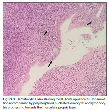
All appendectomy materials were examined histologically
in our hospital within a routine procedure: The specimens
were fixated in 10% formalin for 24 hours and four samples
of five microns thickness were dissected for evaluation. After
macroscopic evaluation, Hematoxylin-eosin stained slides
were examined under a microscope. Clinical diagnoses
and microscopic examination results were recorded. The
cases in which neutrophil infiltration was observed in the
muscularis propria of the appendix were defined as “AA”
histopathologically (Figure 1). The specimens of the two 
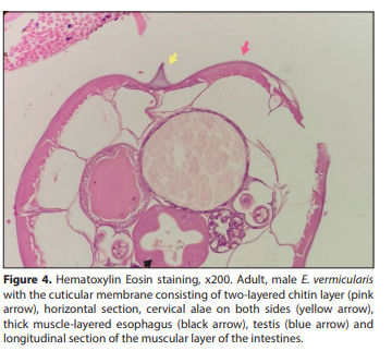
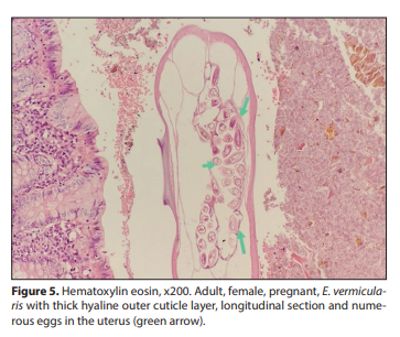
groups were re-evaluated and reported as LH (Figure 2),
congestion, periappendicitis, hemorrhagic appendicitis,
perforated appendicitis, abscess, phlegmonous appendicitis,
gangrenous appendicitis, necrotizing appendicitis and E.
vermicularis with or without peritonitis (Figure 3). The samples
having hemorrhage, perforation, abscess, necrotizing,
phlegmanous and/or gangrenous features were defined to
be “complicated appendicitis.” E. vermicularis cases underwent
additional procedures: Histochemical staining was performed
with Periodic acid- Schiff (PAS) dye to evaluate the cuticular
membrane consisting of a two-layered chitin layer, thick musclelayered esophagus, cervical alae, intestinal walls, uterus and
testicular tissue of the nematode, which were pathognomonic
in histopathological diagnosis of enterobiasis (Figure 4, 5). 
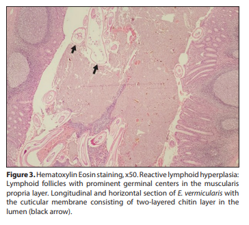
Statistical Analysis
The analyses were performed by statistical package
program software. The data were presented as percentages,
frequencies, medians and minimum-maximum ranges or
mean and standard deviation (SD) by descriptive statistics.
Independent samples t-test was applied for the analyses
of normally distributed continuous variables; whereas
Mann-Whitney U or Kruskal Wallis tests were applied for the
evaluation of abnormally distributed variables with Bonferroni
correction when indicated. Cross-tables with chi-square test
(χ2) and Fisher’s exact test were performed for the evaluation
of categorical variables. Probability factor (p) less than 0.05
was regarded to be statistically significant.
The study was approved by the Ethics Committee of NonInvasive Clinical Research of Amasya University (registration
no: E-76988455-050.01.04-135458).
RESULTS
Mean age of the whole study population was 10.15 ± 3.54
years. The sex ratio of the study and control groups was man/
female: 0.84/ 0.70. The difference in sex was not statistically
significant (p= 0.84). Most of the study group members lived
in rural areas (n= 33; 71.7%), and 31 (67.4%) members of the
control group lived in urban areas, which was statistically
significant (p< 0.0001). The rate of high-income level was
higher in the control group (p= 0.001). Most of the population
in both groups had crowded families (number of household
members ≥6). The frequency of children having their own
room was significantly higher in the control group (p< 0.0001).
Statistical difference was found in the water supply item (p=
0.026); the source of water supply was mains water in 84.8%
(n= 39) of the control group whereas 34.8%; (n= 16) of the
study group had spring water. The frequency of enterobiasis
decreased significantly while the education levels of the
parents increased (p< 0.0001).
The data on symptoms before appendectomy were
obtained from the parental on call surveys. Abdominal pain
and nausea were the most common symptoms in both groups
without a statistical difference (p= 0.39, p= 0.38; respectively).
Pruritus ani presence was significantly higher in the study
group (p< 0.0001). Vomiting, difficulty in defecation and
fever were more frequent in the control group (p< 0.0001;
p< 0.0001; p< 0.0001, respectively). Prevalence of growth
retardation (Body mass index< 5%) was significantly higher in
the study group [39.1%; (n= 18) vs. 10.9%; (n= 5); p< 0.0001].
Approximately half of the patients (n= 50; 53.4%) had
radiological evaluation while the rest went through surgery
on clinical diagnosis. The rate of AA was higher in the study
group (32.6%; n= 15 vs. 19.6%; n= 9), which was statistically
significant (p= 0.001). In addition, only one patient (2.2%) was
diagnosed as “normal” in the control group; but the report of
10.9% (n= 5) of the study group was “normal”, which was a
significant result (p< 0.0001). The appendiceal diameter was
higher and statistically confirmed in the control group (9.05 ±
2.60 vs. 6.50 ± 1.12; p= 0.002).
The average of WBC and neutrophils in CBC were higher
in the control group [14530 ± 5900/mm3
; 10500/mm3
(1500-
29300), respectively]. In addition, median of CRP levels was
1.67 mg/dL (0.5-89.9) in the study group and 12.5 mg/dL
(0.25-172.8) in the control group. Elevation of acute phase
reactants (WBC, neutrophils, CRP) was significantly lower
in the enterobiasis group (p= 0.002; p= 0.005; p< 0.0001,
respectively) whereas the median of eosinophils was higher
(100/mm3 vs. 50/mm3
; p= 0.004). There was no statistical
significance in serum BUN/Cr, bilirubin, sodium and urine
density levels between the groups.
LH, which is thought to be reactive, was the most
frequent histopathological diagnosis in the appendectomy
specimens containing E. vermicularis (n= 27; 58.7%). The rate
of complicated appendicitis was 73.9% (n= 34) in the control
group and LH was not detected. The rates of LH, AA and
complicated appendicitis were different statistically between
the groups (p< 0.0001). Socio-demographic, laboratory,
radiologic and histopathological features of the groups are
summarized in Table 1.
DISCUSSION
E. vermicularis is a nematode requiring a human host and
completing its life cycle usually in the different parts of the
gastrointestinal tract. During its migration, when it visits the
appendix, it may cause “appendiceal colic” and the story ends
with appendectomy. The rate of appendiceal enterobiasis in children of our
hospital during the study period was 1.6%, which was
compatible with the literature. Most of the study group
members were from rural areas and low or intermediate
socioeconomic levels having unsanitary water supply. The
education levels of the parents were lower in the study
group. Vomiting, difficulty in defecation, and fever, which
are symptoms pointing to a salient surgical problem, were
more common in the control group; whereas pruritus ani
and growth retardation prevalences were higher in the study
group, as expected. Lower ultrasonographic appendiceal
diameter, WBC, neutrophil, CRP and higher eosinophil levels
were the features of the study group. The rate of complicated
appendicitis was lower in the study group and LH was the
most frequent histopathological diagnosis.
In a systematic review, a total of 103195 appendicitis cases
were evaluated for E. vermicularis infestation, of which 2983
(2.89%) patients were positive (23). The rate of E. vermicularis
was reported to be 1.5-3% in Türkiye in the same study (23).
Enterobiasis in appendicitis was declared to be 7.69%-80% in
some other reports focusing on the role of parasitic infections
in AA (12,15,24-26). The rate of enterobiasis was inversely
proportional with income levels, which was also compatible
with our results (23).The results on the statistical significance
of sex varied in the literature; the rate of enterobiasis was
higher in girls and boys in different reports (12,19,27). The
prevalence of infestation was higher in rural area residents,
compatible with some reports (27). Low education levels,
crowded household members, and water supply with poor
sanitation conditions are known risk factors in parasitic
infestations as reported in our study (28). The parasite has a
worldwide distribution affecting all population groups and
geographic areas, therefore the results of studies about sociodemographic features may vary (23).
AA is a surgical diagnosis based on clinical suspicion (1).
Pruritus ani is an expected symptom in enterobiasis, as in our
results; however, reports on other gastrointestinal symptoms
such as vomiting, fever, difficulty in defecation have different
conclusions (19,25).This may be because of the evaluation of
different populations. In fact, it is impossible to predict EAAA
just by considering clinical complaints.
Radiological evaluation usually provides confirmation of
AA. Ultrasonography is reported to be more reliable than other
laboratory tests for confirmation or exclusion (29). However,
ultrasonography or other imaging diagnostic techniques are
not reliable to diagnose EAAA (25). In our population, the rate
of AA diagnosis and appendicular diameter were lower in the
EAAA group, compatible with the literature (25).
Elevated WBC, neutrophil and CRP levels establish positive
acute phase response pointing to systemic inflammation.
In our study, compatible with the literature, NEAAA cases
had higher acute phase responses (19,25). Eosinophilia is
expected to be related to parasitic infections, and eosinophil
counts were higher in the EAAA group in our study (19).
However, results of some reports were different, pointing no
relationship with eosinophilia and enterobiasis (25,30). Serum
sodium, BUN/Cr and urine density were analysed to confirm
dehydration, no statistical difference between the groups was found. There was no study focusing on these parameters
in the literature. Serum bilirubin level has been reported
to be a follow up marker of complicated appendicitis in the
literature (4,22). In our study, the results were analysed and
no difference between the groups was detected although
the rate of complicated cases was higher in the control group
(31,32).
The appendix is a lymphoid organ that ends with a blind
space, and therefore, LH is a common finding in appendectomy
specimens, as in this study. Pehlivanoğlu et al. reported LH in
all EAAA cases of their study and concluded that E. vermicularis
was not a reason of AA (33). There are reports pointing to
enterobiasis as the etiologic agent of acute inflammation or
incidental finding, but determining its role in AA is impossible
because of low number of cases and non-homogeneous
study groups and designs (19,34). In this study group, the
rate of AA was 37.9%, so it is also difficult to define the role
of E. vermicularis in AA due to our results. However, increased
intraluminal pressure of appendix may result in appendicitis
by any reason, and enterobiasis may be one of the reasons of
this pathological process.
This study has several limitations: it has a retrospective
nature and small sample size that may not let us come to
definite conclusion. Although our results note the differences
and similarities between EAAA and NEAAA, the data are
based on hospital records and questionnaire answers of the
parents which may have memory bias. Data from physical
examinations were not detailed properly. The surgical
procedures were not mentioned. The sample size is small,
but it is still one of the large paediatric series in the literature
mentioning clinical, hematologic, biochemical, radiologic, and
histopathological data from a holistic point of paediatric view.
Preoperative diagnosis is challenging because AA is one of
the most common surgical problems, but EAAA in these cases
is rare. The history of pruritus ani, lower markers of systemic
inflammation, appendiceal diameter and higher eosinophilia
can be alarming for the surgeon about the parasite. At this
point, medical therapy by anthelminthic drugs and whole
family therapy must be taken in consideration to complete
the therapeutic process.
REFERENCES
1
Dunn James JY. Appendicitis. In: Coran AG, Adzick NS, Krummel TM, et
al. (eds.). Pediatric Surgery. Philadelphia: Elsevier Saunders; 2012:1255-
78. https://doi.org/10.1016/B978-0-323-07255-7.00100-8
2
Gatti S, Lopes R, Cevini C. Intestinal parasitic infections in an institution for the mentally retarded. Ann Trop Med Parasitol 2000;94:453-60.
https://doi.org/10.1080/00034983.2000.11813564
3
Leder K, Weller PF. Enterobiasis (pinworm) and trichuriasis (whipworm)
Uptodate Available from: https://www.uptodate.com. (Accessed date:
May 2024).
4
Ariyarathenam AV, Nachimuthu S, Tang TY, Courtney ED, Harris SA,
Harris AM. Enterobius vermicularis infestation of the appendix and
management at the time of laparoscopic appendectomy: case series
and literature review. Int J Surg 2010;8:466-9. https://doi.org/10.1016/j.
ijsu.2010.06.007
5
Ayudin Ö. Incidental parasitic infestations in surgically removed appendices: a retrospective analysis. Diagn Pathol 2007;2:16. https://doi.
org/10.1186/1746-1596-2-16
6
Arca MJ, Gates RL, Groner JI, Hammond S, Caniano DA. Clinical manifestations of appendiceal pinworms in children: an institutional experience and a review of the literature. Pediatr Surg Int 2004;20:372-5.
https://doi.org/10.1007/s00383-004-1151-5
7
Sousa J, Hawkins R, Shenoy A, Petroze R, Mustafa M, Taylor J, et al. Enterobius vermicularis-associated appendicitis: A 22-year case series and
comprehensive review of the literature. J Pediatr Surg 2022;57:1494-98.
https://doi.org/10.1016/j.jpedsurg.2021.09.038
8
Hasan A, Nafie K, El-Sayed S, Nasr M, Abdulmohaymen A, Baheeg M, et
al. Enterobius vermicularis in appendectomy specimens; Clinicopathological assessment: Cross sectional study. Ann Med Surg 2020;60:168-
72. https://doi.org/10.1016/j.amsu.2020.10.057
9
Still GF. Observations on oxyuris vermicularis in children. Br Med J
1899;1:898-900. https://doi.org/10.1136/bmj.1.1998.898
10
Lamps LW. Infectious causes of appendicitis. Infect Dis Clin North Am
2010;24:995-1018. https://doi.org/10.1016/j.idc.2010.07.012
11
Fleming, CA, Kearney DE, Moriarty P, Redmond HP, Andrews EJ. An evaluation of the relationship between Enterobius vermicularis infestation
and AA in a paediatric population - A retrospective cohort study. Int J
Surg 2015;18:154-8. https://doi.org/10.1016/j.ijsu.2015.02.012
12
Podda M, Gerardi C, Cillara N, Fearnhead N, Gomes CA, Birindelli A,
et al. Antibiotic treatment and appendectomy for uncomplicated
acuteappendicitis in adults and children: a systematic review and
meta-analysis. Ann Surg 2019;270:1028-40. https://doi.org/10.1097/
SLA.0000000000003225
13
Sinniah B, Leopairut J, Neafie RC, Connor DH, Voge M. Enterobiasis:
a histopathological study of 259 patients. Ann Trop Med Parasitol
1991;85:625-35. https://doi.org/10.1080/00034983.1991.11812618
14
Neyzi O, Günöz H, Furman A, Bundan R, Gökçay G, Darendeliler F, et al.
Weight, height, head circumference and body mass index references for
Turkish children. Çocuk Sağlığı ve Hastalıkları Dergisi 2008;51:1-14.
15
Taghipour A, Olfatifar M, Javanmard E, Norouzi M, Mirjalali H, Zali MR.
The neglected role of Enterobius vermicularis in appendicitis: A systematic review and metaanalysis. PLoS ONE 2020;15:e0232143 https://doi.
org/10.1371/journal.pone.0232143
16
Taghipour A, Olfatifar M, Javanmard E, Norouzi M, Mirjalali H, Zali MR.
The neglected role of Enterobius vermicularis in appendicitis: A systematic review and metaanalysis. PLoS ONE 2020;15:e0232143 https://doi.
org/10.1371/journal.pone.0232143
17
Maloney C, Edelman MC, Bolognese AC, Lipskar AM, Rich BS. The impact of pathological criteria on pediatric negative appendectomy
rate. J Pediatr Surg 2019;54:1794-9. https://doi.org/10.1016/j.jpedsurg.2018.10.106
18
Zouari M, Louati H, Abid I, Trabelsi F, Ben Dhaou M, Jallouli M, et al.
Enterobius vermicularis: a cause of abdominal pain mimicking AA in
children: A retrospective cohort study. Arch Iran Med 2018;21:67-72.
19
Gorter RR, van Amstel P, van der Lee JH, van der Voorn P, Bakx R, Heij
HA. Unexpected findings after surgery for suspected appendicitis
rarely change treatment in pediatric patients; results from a cohort
study. J Pediatr Surg 2017;52:1269-72. https://doi.org/10.1016/j.jpedsurg.2017.02.012
20
Lee SE, Lee JH, Ju JW, Lee WJ, Cho SH. Prevalence of Enterobius vermicularis among preschool children in Gimhae-si, Gyeongsangnam-do,
Korea. Korean J Parasitol 2011;49:183-5. https://doi.org/10.3347/
kjp.2011.49.2.183
21
Gunawardena NK, Chandrasena TN, de Silva NR. Prevalence of enterobiasis among primary school children in Ragama, Sri Lanka. Ceylon
Med J 2013;58:106-10. https://doi.org/10.4038/cmj.v58i3.5039
22
Zouari M, Jallouli M, Louati H, Kchaou R, Chtourou R, Kotti A, et al. Predictive value of C-reactive protein, ultrasound and Alvarado score in
AA: a prospective pediatric cohort. Am J Emerg Med. 2016;34:189-92.
https://doi.org/10.1016/j.ajem.2015.10.004
23
Yang Y, Guo C, Gu Z, Hua J, Zhang J, Qian S, et al. The Global Burden
of Appendicitis in 204 Countries and Territories from 1990 to 2019. Clin
Epidemiol 2022;14:1487-99. https://doi.org/10.2147/CLEP.S376665
24
Clark PJ. Utility of eosinophilia as a diagnostic clue in lower abdominal pain in northern Australia: a retrospective case-control
study. Intern Med J 2008;38:278-80. https://doi.org/10.1111/j.1445-
5994.2008.01644.x
25
Zosimas D, Lykoudis PM, Strano G, Burke J, Al-Cerhan E, Shatkar V.
Bilirubin is a specific marker for the diagnosis of AA. Exp Ther Med
2021;22:1056. https://doi.org/10.3892/etm.2021.10490
26
Özen FZ, Celep G. Clinicopathological Assesment in Patients with
Enterobius vermicularis Infection. Turk Parazitol Derg 2023;47:93-9.
https://doi.org/10.4274/tpd.galenos.2023.02418
27
Pehlivanoğlu B, Türk BA, İşler S, Özdas S, Abes M. Findings in appendectomies with Enterobius vermicularis infection: pinworm is not a
cause of appendicitis, Turk Parazitol Derg 2019;43:21-5. https://doi.
org/10.4274/tpd.galenos.2019.6177
28
Chilkar SM, Leelakumar V, Musthyala NB. Enterobius vermicularis infestation with acute perforated suppurative appendicitis in a child: cause
or mere association? J Pediatr Infect Dis 2016;11:19-21. https://doi.
org/10.1055/s-0036-1587598
29
Soyer T. Laboratory Methods in the Diagnosis of Appendicitis. In: Appendicitis and Differential Diagnosis in Children. İzmir: Ege University
Rectorate Publishing; 2018:8-54.
30
Russell RC, Williams NS, Bulstrode CJ. The vermiform appendix. In: Russell RC, Williams NS, Bulstrode CJ, (eds.). Baileyand Love‘s Short Practice
of Surgery. London: Arnold Publishers; 2000:1076-92.
31
Rabah R. Pathology of the appendix in children: an institutional experience and review of the literature. Pediatr Radiol 2007;37:15-20. https://
doi.org/10.1007/s00247-006-0288-x
32
Akbulut S, Tas M, Sogutcu N, Arikanoglu Z, Basbug M, Ulku A, et al.
Unusual histopathological findings in appendectomy specimens: A
retrospective analysis and literature review. World J Gastroenterol
2011;17:1961-70. https://doi.org/10.3748/wjg.v17.i15.1961
33
Juckett G. Common intestinal helminths. Am Family Phys 1995;52:2039-
48
34
Li HM, Zhou CH, Li ZS, Deng ZH, Ruan CW, Zhang QM, et al. Risk factors
for Enterobius vermicularis infection in children in Gaozhou, Guangdong, China. Infect Dis Poverty 2015;4:28. https://doi.org/10.1186/
s40249-015-0058-9